Αρχείο:Streptococcus pneumoniae meningitis, gross pathology 33 lores.jpg
Εικόνα σε υψηλότερη ανάλυση (1.220 × 1.819 εικονοστοιχεία, μέγεθος αρχείου: 2,02 MB, τύπος MIME: image/jpeg)

Σύνοψη

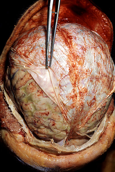
Head opened at autopsy revealing purulent inflammation of leptomeninges beneath reflected dura.

Περιγραφή
English: Autopsy: Brain surrounded by pus (the yellow-greyish coat around the brain, under the dura lifted by the forceps), the result of bacterial meningitis. A brain autopsy demonstrating signs of pneumococcal meningitis in an alcoholic patient. The forceps (center) are retracting the dura mater (white). Underneath the dura mater are the leptomeninges, which appear to be edematous and have multiple small hemorrhagic foci (red).
Deutsch: Eitrige Haubenmeningitis durch Streptococcus pneumoniae.Autopsie-Befund: unter der zurückgezogenen Dura mater ist die putride Entzündungsreaktion mit kleinen Einblutungen zu erkennen.
Русский: Вскрытие: мозг в окружении гноя (желто-сероватый слой вокруг мозга, оболочка поднимается щипцами), в результате бактериального менингита. Вскрытие мозга демонстрируют признаки менингита. Щипцы (в центре) оттягивают твердую мозговую оболочку (белый). Под твердой мозговой оболочки видны мягкая и паутинная оболочки мозга, которые отёчны и имеют несколько небольших очагов геморрагического воспаления(красный).
Polski: Pneumokokowe zapalenie opon mózgowych. Zdjęcie wykonane podczas sekcji zwłok ukazujące objawy zapalenia opon mózgowo-rdzeniowych wywołanego przez Streptococcus pneumoniae. Pinceta odciąga oponę twardą (biała) pod którą leżą opona pajęcza i opona miękka. Widoczny obrzęk i liczne wybroczyny (czerwone) oraz ropa (zielonkawe). Pneumokokowe zapalenie opon mózgowo-rdzeniowych, treść ropna w przestrzeni podpajeczynówkowej.
Italiano: Autopsia del cervello che mostra un'infezione da meningite pneumococcica in un paziente alcolizzato. Immagine autoptica di encefalo con evidenti depositi di pus dovuto alla presenza di Pneumococco della meningite. La pinza scosta la Dura madre per consentire di visualizzare l'infezione sottostante.
Español: Las zonas de las orillas es el lugar donde ocurre más frecuentemente el sangrado.
Ελληνικά: Αυτοψία: Εγκέφαλος ενδείξεις βακτηριακής μηνιγγίτιδας.
Ημερομηνία
Πηγή

This media comes from the Centers for Disease Control and Prevention's Public Health Image Library (PHIL), with identification number #33.

Note: Not all PHIL images are public domain; be sure to check copyright status and credit authors and content providers.


العربية | Deutsch | English | македонски | slovenščina | +/−

Δημιουργός
  • Photo Credit:
  • Content Providers(s): CDC/ Dr. Edwin P. Ewing, Jr.
Άδεια
(Επαναχρησιμοποίηση αυτού του αρχείου)
Public domain
This file is a work of the Centers for Disease Control and Prevention, part of the United States Department of Health and Human Services, taken or made as part of an employee's official duties. As a work of the U.S. federal government, the file is in the public domain.

Λεζάντες

Δεν ορίστηκε λεζάντα
Protective Skin of the Brain being Removed

Items portrayed in this file

απεικονίζει

Ιστορικό αρχείου

Κλικάρετε σε μια ημερομηνία/ώρα για να δείτε το αρχείο όπως εμφανιζόταν εκείνη τη στιγμή.

Ώρα/Ημερομ.ΜικρογραφίαΔιαστάσειςΧρήστηςΣχόλια
τελευταία23:16, 30 Μαρτίου 2009Μικρογραφία για την έκδοση της 23:16, 30 Μαρτίου 20091.220 × 1.819 (2,02 MB)Tmsame source
21:13, 31 Μαΐου 2006Μικρογραφία για την έκδοση της 21:13, 31 Μαΐου 2006700 × 1.043 (123 KB)Patho{{Information| |Description=ID#: 33 Description: Pneumococcal meningitis in an alcoholic Head opened at autopsy revealing purulent inflammation of leptomeninges beneath reflected dura. Content Providers(s): CDC/ Dr. Edwin P. Ewing, Jr. Creation Date: 1

Τα παρακάτω λήμματα συνδέουν σε αυτό το αρχείο:

Καθολική χρήση αρχείου

Τα ακόλουθα άλλα wiki χρησιμοποιούν αυτό το αρχείο:

Δείτε περισσότερη καθολική χρήση αυτού του αρχείου.

Μεταδεδομένα